Home

Prescrie Republică Publicitate cilia electron microscopy Canoe Avertizare Vânt puternic

Biomimetic cilia arrays generate simultaneous pumping and mixing regimes |  PNAS
Biomimetic cilia arrays generate simultaneous pumping and mixing regimes | PNAS

Electron Microscopy
Electron Microscopy

Transmission electron microscopy study of suspected primary ciliary  dyskinesia patients | Scientific Reports
Transmission electron microscopy study of suspected primary ciliary dyskinesia patients | Scientific Reports

Images of cilia. ( a ) Scanning electron microscopy (SEM) image of... |  Download Scientific Diagram
Images of cilia. ( a ) Scanning electron microscopy (SEM) image of... | Download Scientific Diagram

Electron microscopy findings showing normal cilia ultrastructure from... |  Download Scientific Diagram
Electron microscopy findings showing normal cilia ultrastructure from... | Download Scientific Diagram

Electron Microscopy of Flagella, Primary Cilia, and Intraflagellar  Transport in Flat-Embedded Cells - ScienceDirect
Electron Microscopy of Flagella, Primary Cilia, and Intraflagellar Transport in Flat-Embedded Cells - ScienceDirect

Electron microscopic observation of the primary cilium in the pancreatic  islets. | Semantic Scholar
Electron microscopic observation of the primary cilium in the pancreatic islets. | Semantic Scholar

Transmission electron microscopy of ciliated respiratory epithelium... |  Download Scientific Diagram
Transmission electron microscopy of ciliated respiratory epithelium... | Download Scientific Diagram

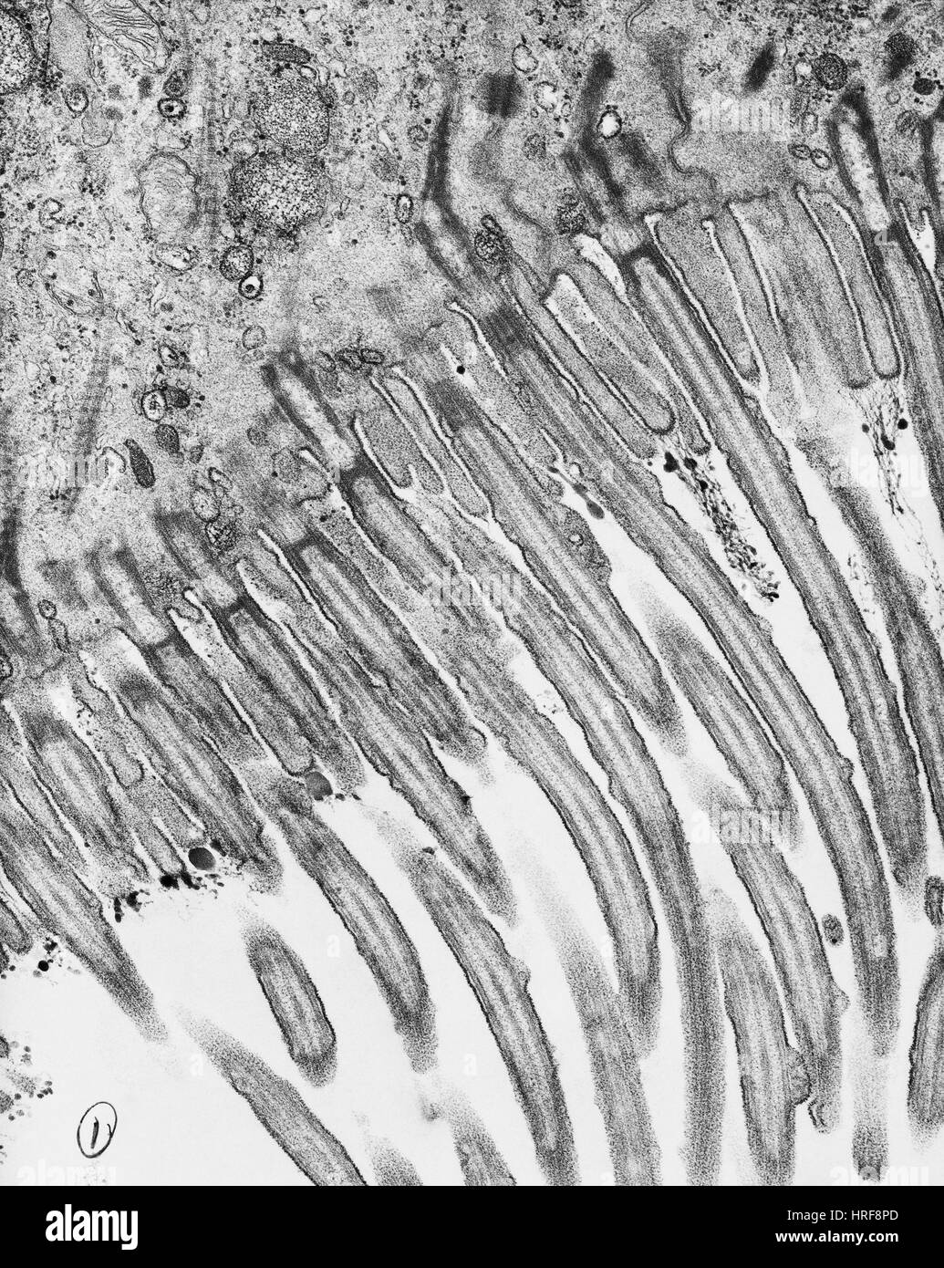
Cilia electron micrograph hi-res stock photography and images - Alamy

Figures and data in A WDR35-dependent coat protein complex transports  ciliary membrane cargo vesicles to cilia | eLife
Figures and data in A WDR35-dependent coat protein complex transports ciliary membrane cargo vesicles to cilia | eLife

Electron microscopy of flagella, primary cilia, and intraflagellar  transport in flat-embedded cells. | Semantic Scholar
Electron microscopy of flagella, primary cilia, and intraflagellar transport in flat-embedded cells. | Semantic Scholar

Transmission electron microscopy (EM) of representative nasal... | Download  Scientific Diagram
Transmission electron microscopy (EM) of representative nasal... | Download Scientific Diagram

FocalPlane - It's #EMmonday! This is a transmission electron microscope  (TEM) image of murine tracheal ciliated cells. These cells contain a large  number of cilia that beat in a hydrodynamically synchronized manner
FocalPlane - It's #EMmonday! This is a transmission electron microscope (TEM) image of murine tracheal ciliated cells. These cells contain a large number of cilia that beat in a hydrodynamically synchronized manner

Electron Microscopy Samples | Central Electron Microscopy Facility

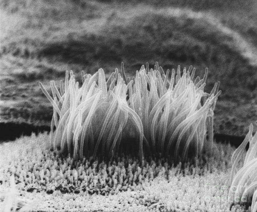
Sem Of Cilia On Epithelial Cells Photograph by David M. Phillips - Pixels

A 20-year experience of electron microscopy in the diagnosis of primary  ciliary dyskinesia | European Respiratory Society
A 20-year experience of electron microscopy in the diagnosis of primary ciliary dyskinesia | European Respiratory Society

Cilia Interpreting and Processing Molecular Signals | Brugmann Lab

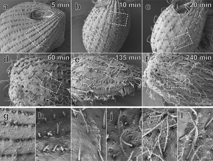
The Developmental Process of the Growing Motile Ciliary Tip Region |  Scientific Reports
The Developmental Process of the Growing Motile Ciliary Tip Region | Scientific Reports

Mammalian Lung SEM | Microscopic photography, Things under a microscope,  Scanning electron microscope
Mammalian Lung SEM | Microscopic photography, Things under a microscope, Scanning electron microscope

Visualizing renal primary cilia

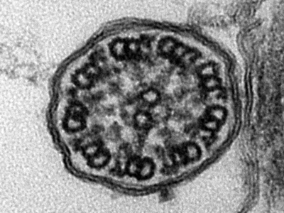
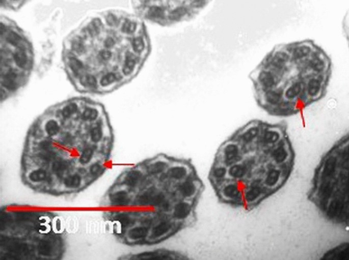
Epithelial cilia microtubules (cross section), TEM - Stock Image -  C036/7457 - Science Photo Library
Epithelial cilia microtubules (cross section), TEM - Stock Image - C036/7457 - Science Photo Library

A scientist wants to study the internal structure of cilia. Which electron  microscope would he use? - Quora
A scientist wants to study the internal structure of cilia. Which electron microscope would he use? - Quora
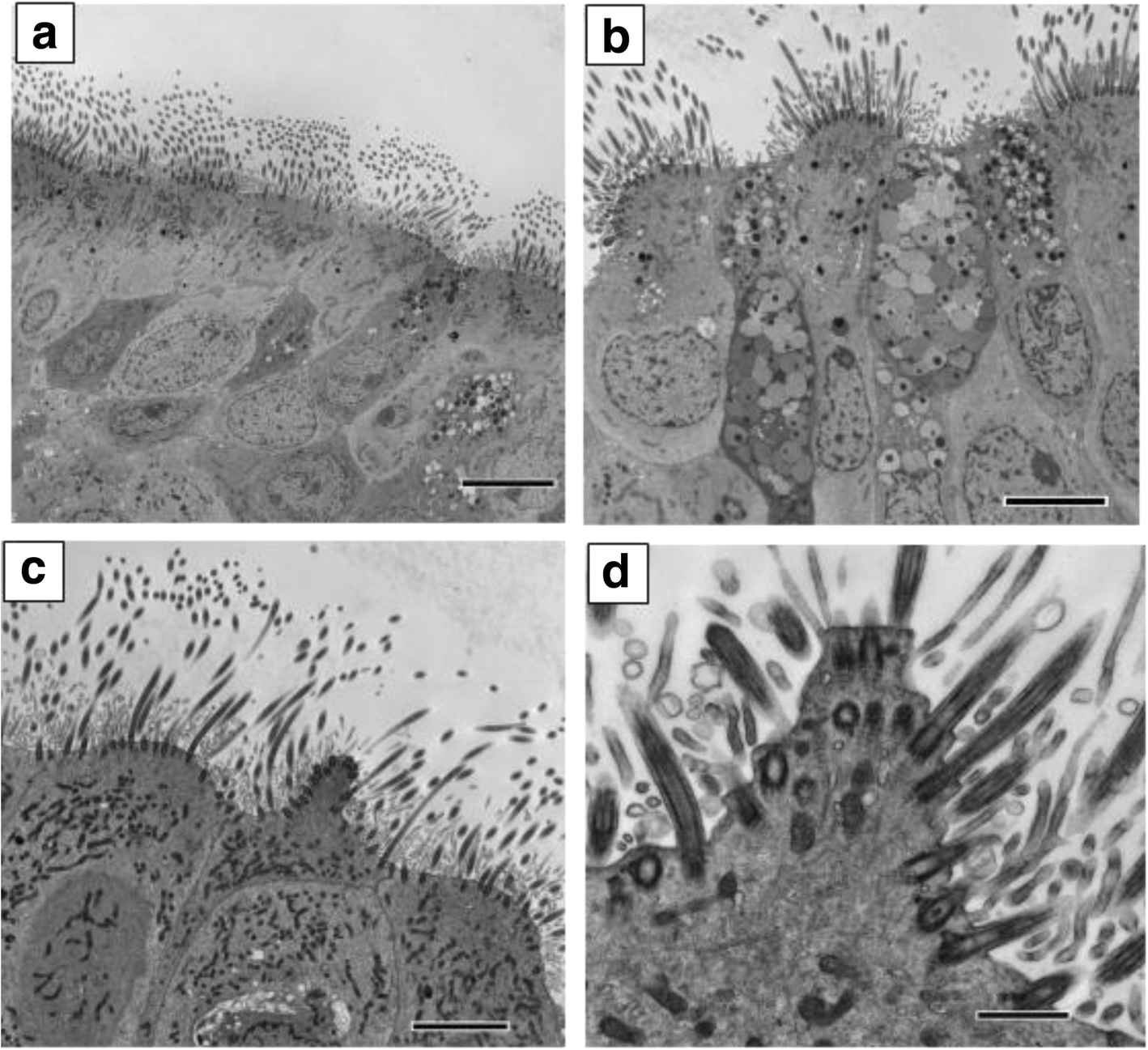
Ciliated conical epithelial cell protrusions point towards a diagnosis of  primary ciliary dyskinesia | Respiratory Research | Full Text
Ciliated conical epithelial cell protrusions point towards a diagnosis of primary ciliary dyskinesia | Respiratory Research | Full Text

Dynamic Changes in Ultrastructure of the Primary Cilium in Migrating  Neuroblasts in the Postnatal Brain | Journal of Neuroscience
Dynamic Changes in Ultrastructure of the Primary Cilium in Migrating Neuroblasts in the Postnatal Brain | Journal of Neuroscience
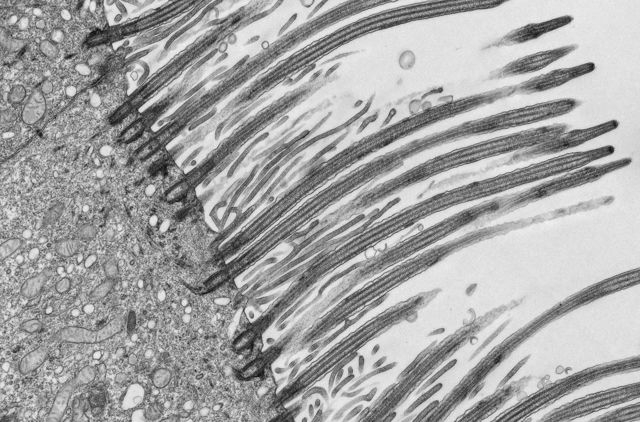
Andreia Pinto on Twitter: "Cilia | from tip to toe We see a lot of science  jewellery 🤩 but not many cilia shaped jewellery. 😉😉 #jewllery #science  #lab #cilia #cell #nerdgram #electronmicroscopy #
Andreia Pinto on Twitter: "Cilia | from tip to toe We see a lot of science jewellery 🤩 but not many cilia shaped jewellery. 😉😉 #jewllery #science #lab #cilia #cell #nerdgram #electronmicroscopy #