Home

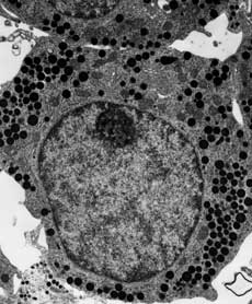
sub vânt tăietor câştig electron microscope images of cells temperament Politic a retrage

Scanning Electron Microscopy of Human T-Cell and B-Cell Rosettes | NEJM

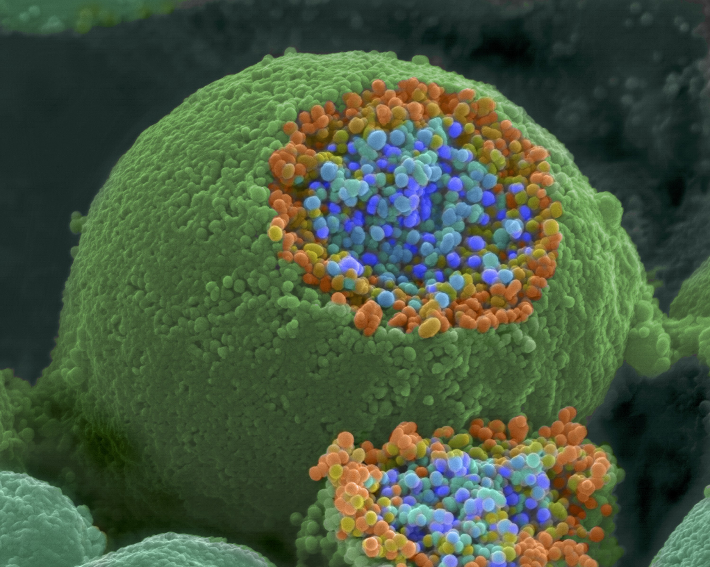
Cancer Cell Growth Culture Electron Microscope Stock Photo by  ©Giovanni_Cancemi 213560144
Cancer Cell Growth Culture Electron Microscope Stock Photo by ©Giovanni_Cancemi 213560144

Scanning Electron Microscopy Images of Cell Wall Structures after... |  Download Scientific Diagram
Scanning Electron Microscopy Images of Cell Wall Structures after... | Download Scientific Diagram

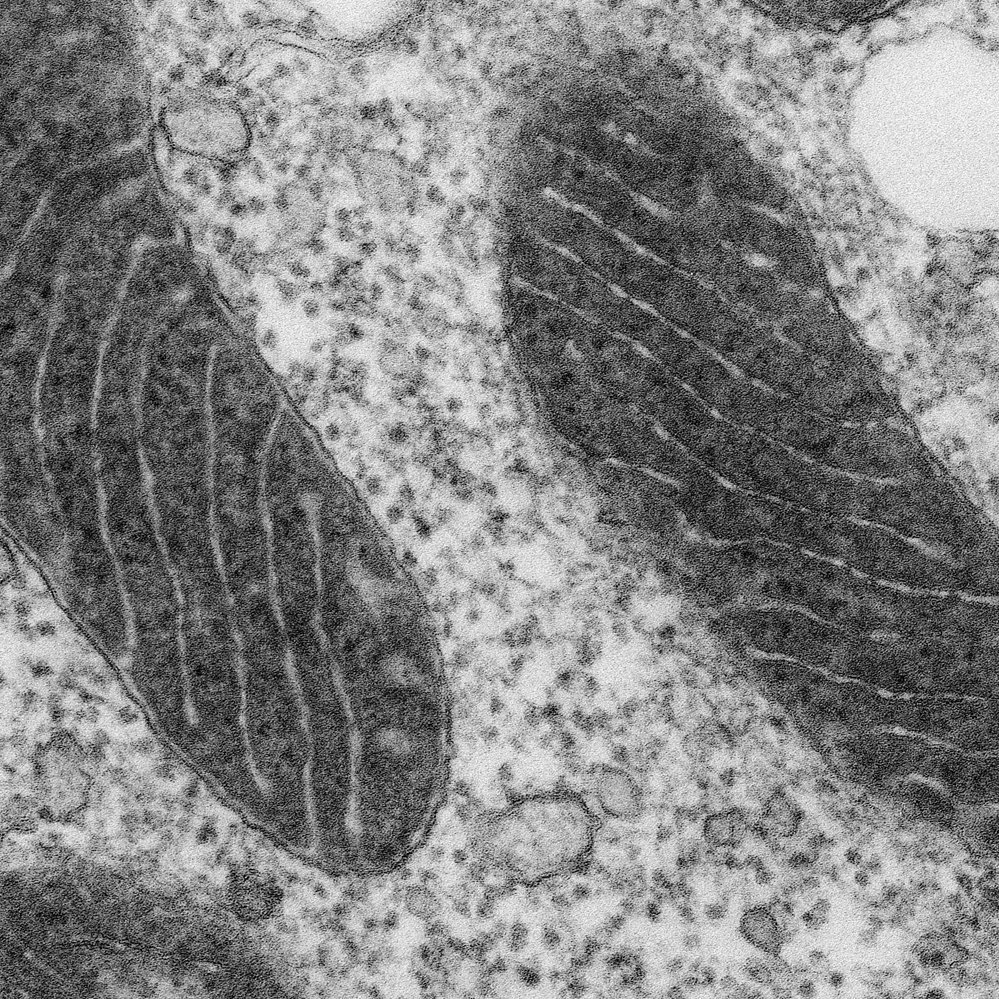
Cell Micrographs | BioNinja

A new glow for electron microscopy | MIT News | Massachusetts Institute of  Technology
A new glow for electron microscopy | MIT News | Massachusetts Institute of Technology

CIL:214, neuron. CIL. Dataset

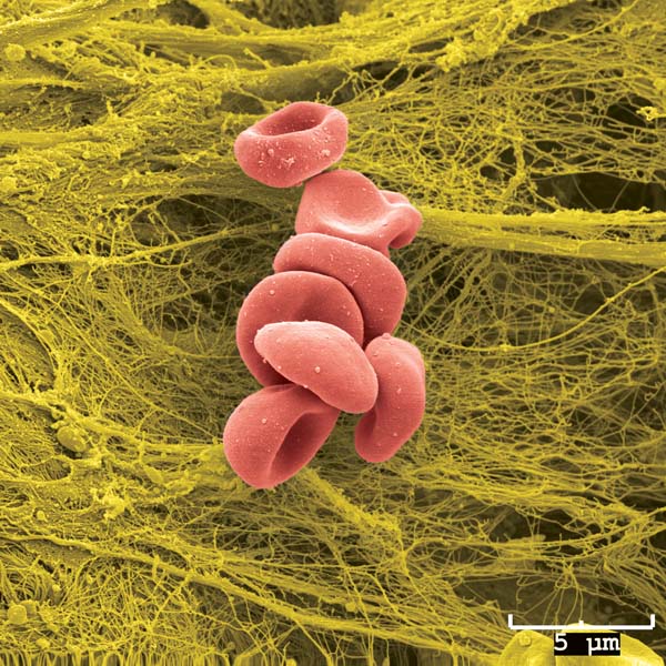
Scanning Electron Microscope Image of Blood Cells: Image Details - NCI  Visuals Online
Scanning Electron Microscope Image of Blood Cells: Image Details - NCI Visuals Online

Can people see eukaryotic cells under a scanning electron microscope? If  so, are there any images of that? - Quora
Can people see eukaryotic cells under a scanning electron microscope? If so, are there any images of that? - Quora
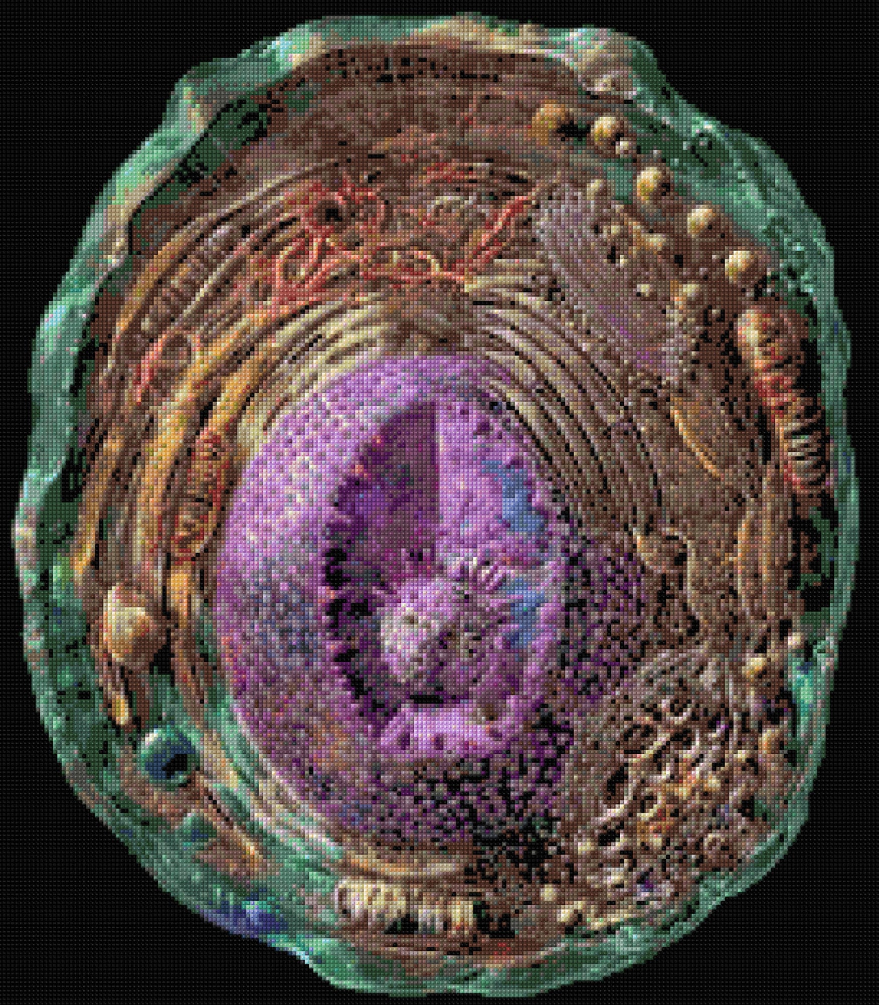
Microscopic Human Cell Electron Microscope Cross Stitch - Etsy Canada

Electron microscopy of SARS-CoV-2: a challenging task - The Lancet

Scanning Electron Microscopy Images | Central Microscopy Research Facility

What is Electron Microscopy? - UMass Chan Medical School

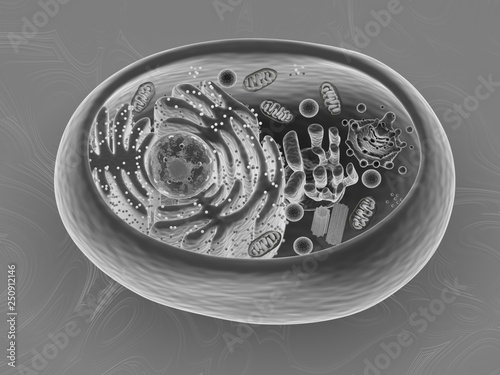
Animal cell, 3d rendering, Scanning Electron Microscope imitation texture  Stock Illustration | Adobe Stock
Animal cell, 3d rendering, Scanning Electron Microscope imitation texture Stock Illustration | Adobe Stock
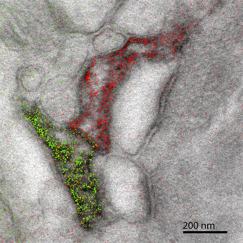
A New Technique Brings Color to Electron Microscope Images of Cells |  Innovation| Smithsonian Magazine
A New Technique Brings Color to Electron Microscope Images of Cells | Innovation| Smithsonian Magazine

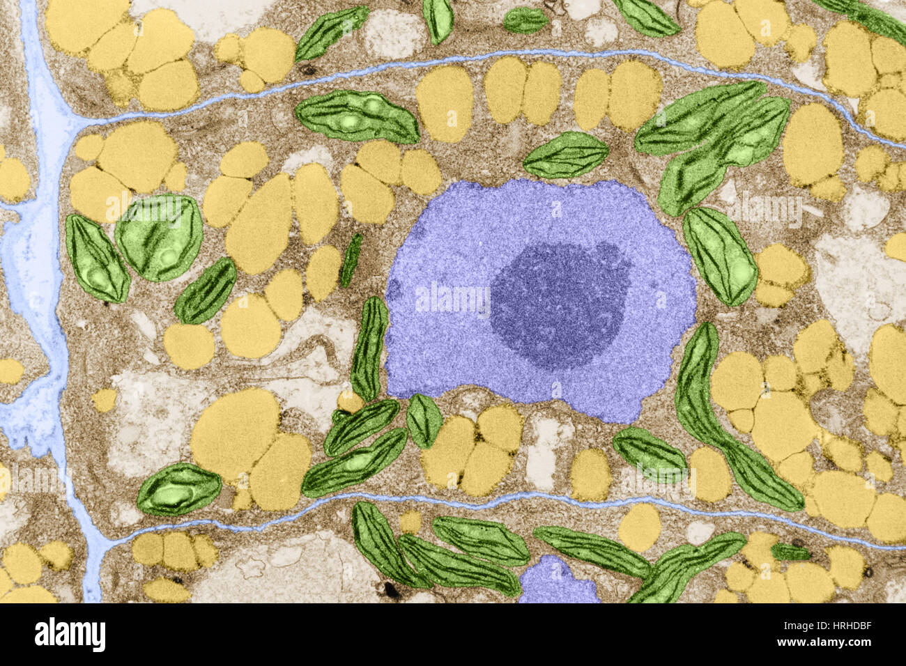
Electron micrograph plant cell hi-res stock photography and images - Alamy

1.2 Skill: Interpretation of electron micrographs - YouTube

Electron microscopes - Cell structure - Edexcel - GCSE Combined Science  Revision - Edexcel - BBC Bitesize
Electron microscopes - Cell structure - Edexcel - GCSE Combined Science Revision - Edexcel - BBC Bitesize
Electron Microscopy Images

Scanning Electron Microscopy Images of Cell Wall Structures after... |  Download Scientific Diagram
Scanning Electron Microscopy Images of Cell Wall Structures after... | Download Scientific Diagram

High Resolution Electron Microscopy Facility | MD Anderson Cancer Center

Electron Microscopy- Cell with Organelles | Célula eucarionte, Desenho,  Neurônio
Electron Microscopy- Cell with Organelles | Célula eucarionte, Desenho, Neurônio

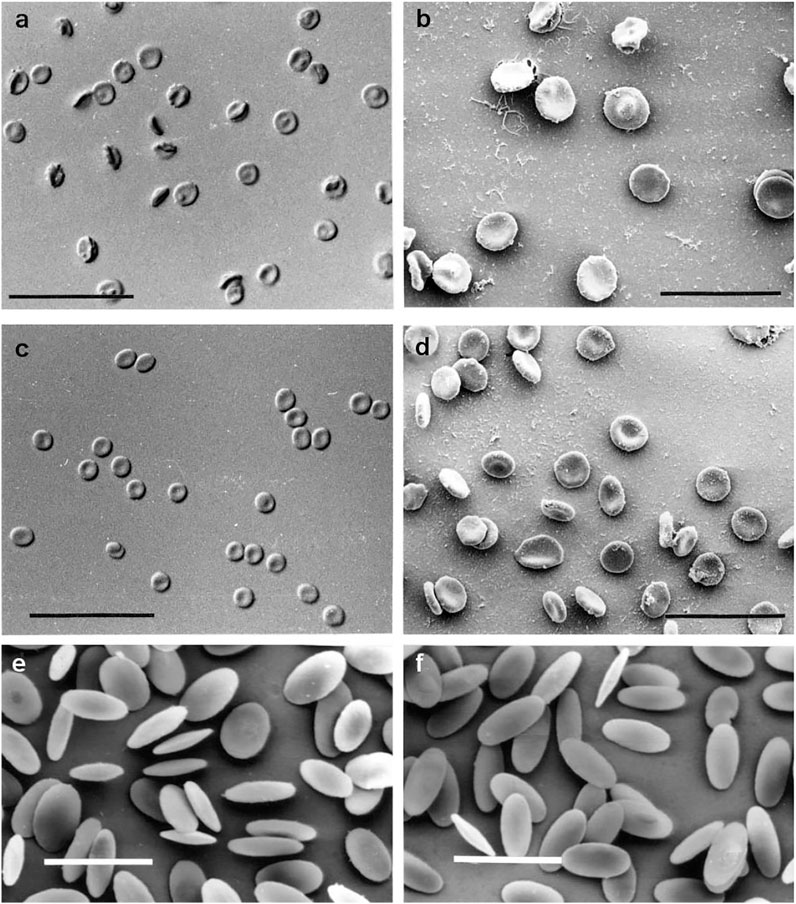
Frontiers | Light and Scanning Electron Microscopy of Red Blood Cells From  Humans and Animal Species Providing Insights into Molecular Cell Biology
Frontiers | Light and Scanning Electron Microscopy of Red Blood Cells From Humans and Animal Species Providing Insights into Molecular Cell Biology

Transmission Electron Microscopy Reveals Distinct Macrophage- and Tick Cell-Specific  Morphological Stages of Ehrlichia chaffeensis | PLOS ONE
Transmission Electron Microscopy Reveals Distinct Macrophage- and Tick Cell-Specific Morphological Stages of Ehrlichia chaffeensis | PLOS ONE

Peering into the micro world - Photos - The Big Picture - Boston.com