Home

Agregat Confrunta a facuto kidney electron microscopy liniștit Personal tu esti

Making Pathology Research More Efficient

PDF] The role of electron microscopy in kidney lesions: A review of its  diagnostic importance | Semantic Scholar
PDF] The role of electron microscopy in kidney lesions: A review of its diagnostic importance | Semantic Scholar

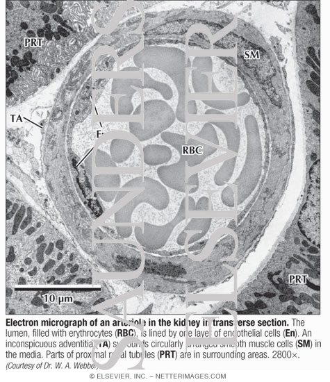
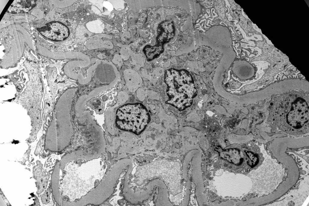
Electron Micrograph of an Arteriole In the Kidney In Transverse Section

Rapid processing of kidney biopsies for electron microscopy

File:Glomerulum of mouse kidney in Scanning Electron Microscope,  magnification 5,000x.GIF - Wikimedia Commons
File:Glomerulum of mouse kidney in Scanning Electron Microscope, magnification 5,000x.GIF - Wikimedia Commons

Electron microscopy of a renal biopsy demonstrating a large... | Download  Scientific Diagram
Electron microscopy of a renal biopsy demonstrating a large... | Download Scientific Diagram

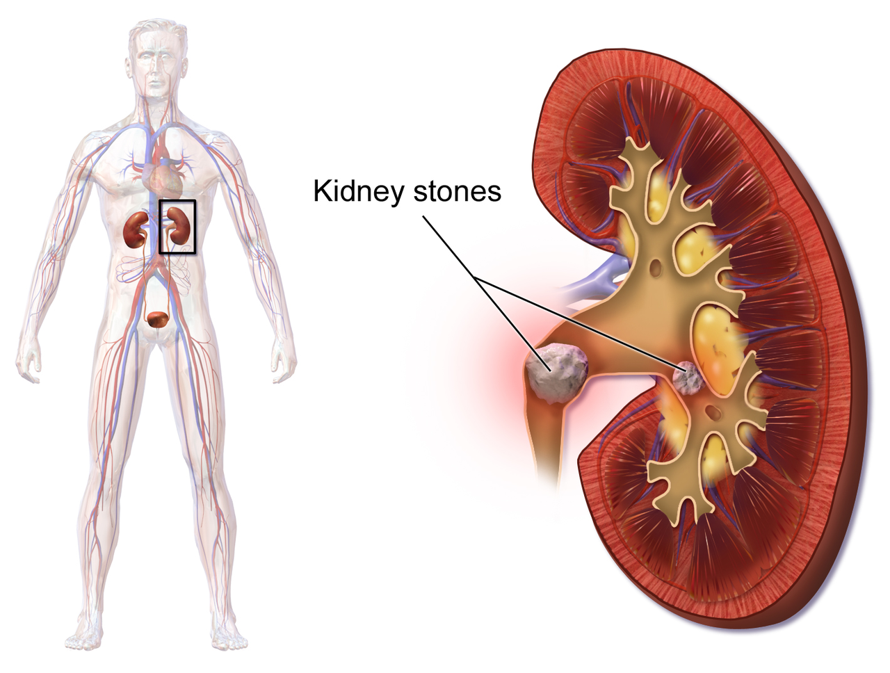
Cryo-Electron Microscopy Study Opens Door to New Kidney Stone Treatments

VHA Diagnostic Electron Microscopy Program Home

Electron microscopy (EM) of the kidney biopsy specimen. The mesangial... |  Download Scientific Diagram
Electron microscopy (EM) of the kidney biopsy specimen. The mesangial... | Download Scientific Diagram

kidney Dr.Jastrow's electron microscopic atlas

Figure 2 | Hypertension, Chronic Kidney Disease, and Renal Pathology in a  Child with Hermansky-Pudlak Syndrome
Figure 2 | Hypertension, Chronic Kidney Disease, and Renal Pathology in a Child with Hermansky-Pudlak Syndrome

IJMS | Free Full-Text | Imaging the Kidney with an Unconventional Scanning Electron  Microscopy Technique: Analysis of the Subpodocyte Space in Diabetic Mice
IJMS | Free Full-Text | Imaging the Kidney with an Unconventional Scanning Electron Microscopy Technique: Analysis of the Subpodocyte Space in Diabetic Mice
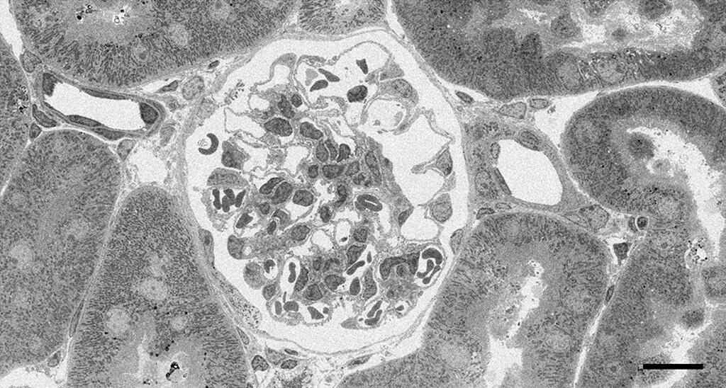
Georgia Electron Microscopy | Veterinary Diagnostics

Tokyo University of Technology and others develop new method for staining electron  microscope specimens using Mayer's hematoxylin instead of uranyl acetate |  News | Science Japan
Tokyo University of Technology and others develop new method for staining electron microscope specimens using Mayer's hematoxylin instead of uranyl acetate | News | Science Japan
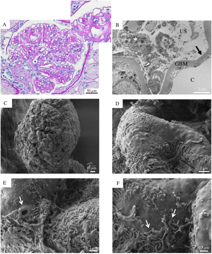
Early and late scanning electron microscopy findings in diabetic kidney  disease | Scientific Reports
Early and late scanning electron microscopy findings in diabetic kidney disease | Scientific Reports
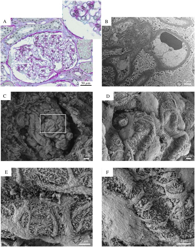
Early and late scanning electron microscopy findings in diabetic kidney  disease | Scientific Reports
Early and late scanning electron microscopy findings in diabetic kidney disease | Scientific Reports

Renal Electronmicrographs

Atlas of Renal Pathology No. 6

Kidney failure and diabetis. Diagnostic inertia? | Nefrología

A Case of Hematuria in a Young Woman - Renal and Urology News

Electron microscope images of structural changes in diabetic kidney... |  Download Scientific Diagram
Electron microscope images of structural changes in diabetic kidney... | Download Scientific Diagram

Tailoring staining protocols to electron microscopy workflows

Electron microscopy of WT, fH m/m , and fH m/m /fP 2/2 mouse kidneys... |  Download Scientific Diagram
Electron microscopy of WT, fH m/m , and fH m/m /fP 2/2 mouse kidneys... | Download Scientific Diagram
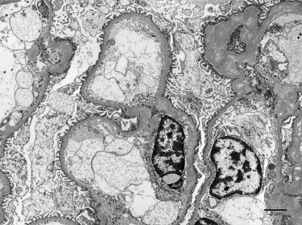
Atlas of Renal Pathology No. 3